Home
/ Anatomy Of The Back Of The Neck Muscles - Soft Tissue Pain in the Neck - The Buxton Osteopathy Clinic : Intermediate back muscles and c.
Anatomy Of The Back Of The Neck Muscles - Soft Tissue Pain in the Neck - The Buxton Osteopathy Clinic : Intermediate back muscles and c.
Anatomy Of The Back Of The Neck Muscles - Soft Tissue Pain in the Neck - The Buxton Osteopathy Clinic : Intermediate back muscles and c.. Understanding the anatomy and function of your back muscles can help you determine if (and when) you may need professional medical care if you are having a problem with your back. Still, many individuals pay far too little attention to them. The platysma subcutaneous muscle of the neck (platysma) extends from the chin to the pectoral region. The extensors and rotators of the head and neck: The splenius capitis and cervicis (spinotransversales muscles).
From the sides and the back of the neck, the splenius capitis inserts onto the head region, and the splenius. Explanations spinoscapular and spinohumeral muscles the extrinsic back muscles are also referred to as secondary back muscles. Some beginners aren't even aware that the neck. And the activity at our anterior neck muscles is connected in a wonderful way to the activity of. The posterior muscles of the neck are primarily concerned with head movements, like extension.
Dropped Head Syndrome | eOrthopod.com from www.eorthopod.com Four groups of muscles in neck. And the activity at our anterior neck muscles is connected in a wonderful way to the activity of. Click now to learn more at kenhub! This article provides an overview of the neck muscles, their anatomy, origins, insertions, actions, and innervation. Of all the anatomical areas, the neck has the highest proportion of muscles per surface, which is logical considering that these muscles must keep the weight of the head in place, provide mobility and protect vascular and nervous structures, as well as the upper digestive and aerial route. Exercising the back helps reduce muscle stiffness and strain by keeping the connective fibers of ligaments and tendons flexible. This muscle inserts onto the back of the skull just behind the mastoid process and it actually inserts on the mastoid process as those are the muscles of the posterior triangle of the neck. They are divided into three groups, as shown below.
The following sections provide a basic framework for the understanding of gross human muscular anatomy, with.
Extensor musculature of the posterior neck is often tight because it is so often used. The physicians originally studying human anatomy thought the skull looked like an helmet. There are a number of specific muscles within the back anatomy, and its important to take a quick look at all of them to see how you can target them efficiently and develop a strong back. The sternocleidomastoid muscle, or scm, is one of the larger and more superficial muscles in the neck, making it an important and easily identifiable anatomical landmark. Educational video describing the muscle anatomy of the neck. Understanding the anatomy and function of your back muscles can help you determine if (and when) you may need professional medical care if you are having a problem with your back. Muscles of the head & neck | anatomy model. Human muscle system, the muscles of the human body that work the skeletal system, that are under voluntary control, and that are concerned with movement, posture, and balance. Learn about anatomy neck muscles with free interactive flashcards. Neck muscles, like those of the lower back, are often ignored. The trapezius muscle actually considered to be just as much of a muscle related to the back as it is the neck. Muscle spasming of the neck is likely the most common musculoskeletal complaint that exists. From the sides and the back of the neck, the splenius capitis inserts onto the head region, and the splenius.
This is a table of skeletal muscles of the human anatomy. The back muscles can be three types. The suboccipital muscles act to rotate the head and extend the neck. C7 powers the triceps muscle on the back of your upper arms and transmits sensation along the back of the arms, and down to the middle finger. Indeed it is rare to find a client/patient whose neck is not tight.
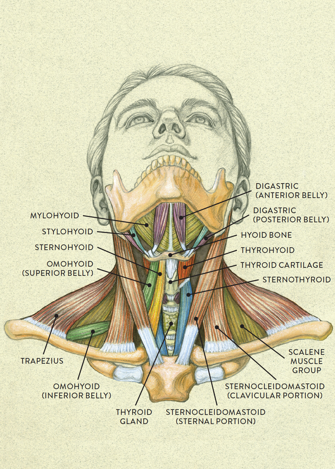
Anterior view of head tilting back from schoolbag.info They are divided into three groups, as shown below. The posterior muscles of the neck are primarily concerned with head movements, like extension. This muscle inserts onto the back of the skull just behind the mastoid process and it actually inserts on the mastoid process as those are the muscles of the posterior triangle of the neck. Click now to learn more at kenhub! Some bodybuilders become so focused on developing the more prominent muscles that they fail to work on subtle muscles, such as those of the neck, that would complete their development. There are a number of specific muscles within the back anatomy, and its important to take a quick look at all of them to see how you can target them efficiently and develop a strong back. Working in pairs on the. The thyrohyoid is a quadrilateral muscle located in the muscular triangle of the neck.
The posterior muscles of the neck are primarily concerned with head movements, like extension.
The sternocleidomastoid muscle, or scm, is one of the larger and more superficial muscles in the neck, making it an important and easily identifiable anatomical landmark. Injuries of the intrinsic back muscles often occur while using improper lifting technique. To borrow a common phrase of renowned author and. The human back is the large posterior area of the human body, rising from the top of the buttocks, to the back of the neck and shoulders. Click now to learn more at kenhub! Exercising the back helps reduce muscle stiffness and strain by keeping the connective fibers of ligaments and tendons flexible. The thyrohyoid is a quadrilateral muscle located in the muscular triangle of the neck. The image below to shows all the major back muscles (as well as some neck muscles): C7 powers the triceps muscle on the back of your upper arms and transmits sensation along the back of the arms, and down to the middle finger. Border of mandible and skin, and is attached to superficial fascia covering pectoralis major and deltoid muscles inferiorly. Extensor musculature of the posterior neck is often tight because it is so often used. Since the all the back muscles originate in embryo (fetus) form by locations… they consist of: Understanding the anatomy and function of your back muscles can help you determine if (and when) you may need professional medical care if you are having a problem with your back.
Since the all the back muscles originate in embryo (fetus) form by locations… they consist of: Explanations spinoscapular and spinohumeral muscles the extrinsic back muscles are also referred to as secondary back muscles. The human back is the large posterior area of the human body, rising from the top of the buttocks, to the back of the neck and shoulders. The classic computer position shortens the posterior (back) neck muscles, making them tight and, over time, possibly shorter. Understanding the anatomy of your cervical spine and the vital nerves it contains should motivate you to adopt behaviors that help prevent neck injury and.
Beautiful illustration of the deep and superficial ... from i.pinimg.com The image below to shows all the significant back muscles (in addition to some neck muscles) This is a table of skeletal muscles of the human anatomy. The splenius capitis and cervicis (spinotransversales muscles). Obliquus capitis superior also extends from the occiput to c1 while obliquus. The back muscles stabilize and move the vertebral column, and are grouped according to the lengths and direction of the fascicles. The neck muscles, including the sternocleidomastoid and the trapezius, are responsible for the gross motor movement in the muscular system of the head and neck. The sternocleidomastoid muscle, or scm, is one of the larger and more superficial muscles in the neck, making it an important and easily identifiable anatomical landmark. This article covers the anatomy of the deep muscles of the back, including their function, blood supply, innervation, origin and insertion.
Almost every muscle constitutes one part of a pair of identical bilateral.
Educational video describing the muscle anatomy of the neck. The splenius capitis and cervicis (spinotransversales muscles). Of all the anatomical areas, the neck has the highest proportion of muscles per surface, which is logical considering that these muscles must keep the weight of the head in place, provide mobility and protect vascular and nervous structures, as well as the upper digestive and aerial route. The suboccipital muscles act to rotate the head and extend the neck. They move the head in every direction, pulling the skull and jaw towards the shoulders, spine, and scapula. Its attachment to the frontal and occipital bellies (muscles on the brow at the front and on the upper back of the head). Table of contents the extrinsic muscles of the back: They are divided into three groups, as shown below. Border of mandible and skin, and is attached to superficial fascia covering pectoralis major and deltoid muscles inferiorly. The sternocleidomastoid muscle, or scm, is one of the larger and more superficial muscles in the neck, making it an important and easily identifiable anatomical landmark. Working in pairs on the. There are a number of specific muscles within the back anatomy, and its important to take a quick look at all of them to see how you can target them efficiently and develop a strong back. Explanations spinoscapular and spinohumeral muscles the extrinsic back muscles are also referred to as secondary back muscles.
The posterior muscles of the neck are primarily concerned with head movements, like extension anatomy of back of neck. The image below to shows all the significant back muscles (in addition to some neck muscles)